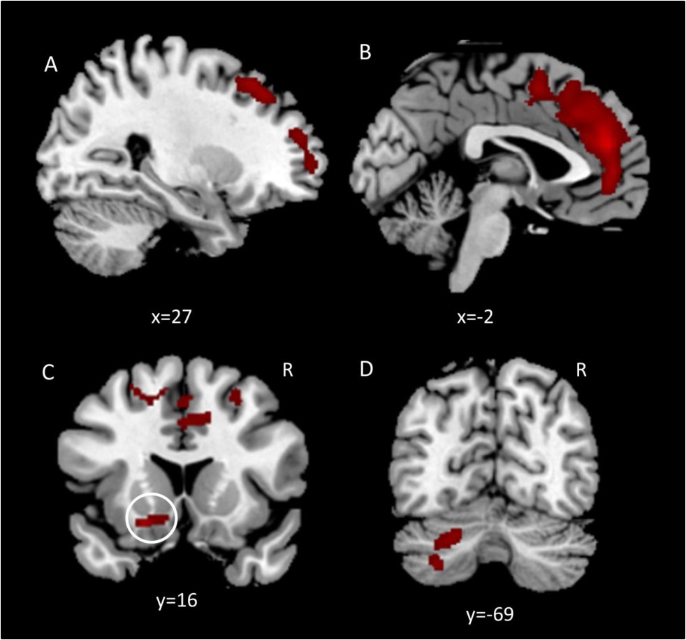
Fig. 2. Clusters showing significant time by group interaction in a whole brain VBM contrasts (p<0.05 (FWE corrected), k > 200, corrected for non-stationary smoothness). Panel A: Right middle frontal gyrus BA 8 (Cluster 5), BA 9/10 (Cluster 2); Panel B: Anterior cingulate cortex (ACC (Cluster 1); Panel C: Nucleus accumbens (Cluster 7, Panel C); Panel D: Cerebellar crus I/II and cerebellar VI (Cluster 4, Cluster 6)